


Immunoprecipitation (IP) is a method to isolate a specific antigen from a mixture, using the antigen-antibody interaction. Antigens isolated by IP are analyzed by SDS-PAGE or Western blotting.
The principle
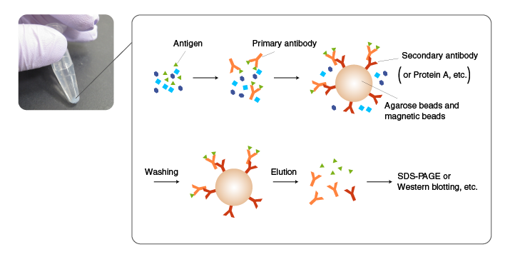
In IP, an antibody is added first to a mixture containing an antigen, and incubated to allow antigen-antibody complexes to form.
Subsequently, the antigen-antibody complexes are incubated with an immobilized antibody against the primary antibody (secondary antibody) or with protein A/G-coated beads to allow them to absorb the complexes.
The beads are then thoroughly washed, and the antigen is eluted from the beads by an acidic solution or SDS.
If suitable antibody is not available, the target molecule is fused to a His tag or other tags by recombinant DNA techniques, and immunoprecipitated using an antibody to the tag (pull-down assay).
[Related topics] Pull-down assay experiments using Tagged protein purification kits

Antibodies raised against synthetic peptides and recombinant proteins often work well in Western blotting but may not bind the antigens in their native conformation in solution.
When using commercial antibodies, select the ones that are suitable for IP according to product information. Also recognize the properties of both the antibody and antigen in the literature and product information.
Tips for efficient IP
The diagram on the right illustrates the structures of antigen-antibody complexes formed with a monoclonal versus polyclonal antibody as the primary antibody.
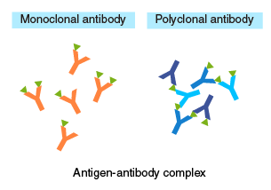 When using a monoclonal antibody as the primary antibody, adjust the concentration so that:
[secondary antibody] > [primary antibody] > [antigen].
An excess of primary antibody, relative to the secondary antibody, may compete with antigen-antibody complexes for the secondary antibody, resulting in a lower yield of recovery.
When using a polyclonal antibody, an excess of primary antibody, relative to the antigen, will prevent the formation of oligomeric complexes. Therefore, these concentrations should be optimized by testing different ratios.
| Monoclonal antibody | Polyclonal antibody | |
|---|---|---|
| Background | Background is low with an appropriate antibody because the antibody recognizes a single antigen. | Background may be high if some of the antibody molecules have a low specificity and bind to proteins other than the target protein. |
| Binding affinity | High-affinity monoclonal antibody (dissociation constant Kd<10-8 M) should be used because low affinity antibody may not form an antigen-antibody complex in solution. | Even if the affinity of individual antibody molecules is low, oligomeric antigen-antibody complexes are formed easily due to the multivalent binding. |
| Stability of antigen-antibody complexes | If the binding affinity of an antibody is low, simultaneously using several high-specificity monoclonal antibodies will allow multivalent binding, resulting in stable antigen-antibody complexes. | Stable oligomeric complexes are formed because reaction between a polyclonal antibody and an antigen is multivalent. |
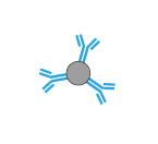
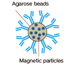
Beads for immobilization
Agarose beads and magnetic beads are commonly used.
Agarose beads have a porous, mesh-like structure, and antibodies can diffuse and bind to the internal matrix of the beads, which provides high binding capacity.
Magnetic beads are simple spheres, providing ease of handling and short processing time. With appropriate coating, background can be reduced. However, binding capacity may not be high enough for some applications, and the cost is higher than the alternative using agarose beads.
Magnetic agarose beads provide ease of handling (when used with a magnet) and a high binding capacity.
| Agarose beads | Magnetic beads | Magnetic agarose beads | |
|---|---|---|---|
| Diameter (MBL products) |
Approx. 100 µm | Approx. 1.6 µm | Approx. 50 µm |
| Diagram of the bead structure and antibody coat | 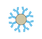 |
 |
 |
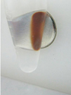
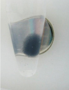
| Actual images |  |
 |
 |
| IgG binding capacity per matrix | High | Moderate | High |
| Likelihood of sample loss | Sample loss may occur during the washing step. | Sample loss is negligible because the beads are pelleted using a magnet. | Sample loss is minimal because the beads are pelleted using a magnet. |
| Centrifugation | Necessary | Unnecessary | Unnecessary |
| Magnetic rack | Unnecessary | Necessary | Necessary |
| Visibility of the beads | Poor | Excellent | Excellent |
| Other features | Inexpensive | Easy to disperse; useful for screening, etc. | Less expensive and provides a higher yield than magnetic beads. |
[Related topics] Smart-IP tag-antibodies conjugated with magnetic beads or magnetic agarose
Detergents
Appropriate concentrations of salt and non-ionic detergent are commonly used in IP to reduce non-specific binding (protein-protein and protein-bead interactions). Pilot experiments should be performed with detergents, which may reduce the affinity of the antibody, especially when using a monoclonal antibody.
Protease inhibitors
The target protein and antibody are subject to degradation by protein-degrading enzymes (proteases) in samples such as cell lysates and tissue extracts. To prevent proteolytic degradation, protease inhibitors are included. When the type of proteases in samples are known or predicted, specific inhibitors are used. When proteases are unknown, a combination of multiple small molecule inhibitors, such as PMSF and EDTA, are used.
Elution condition
In most applications, proteins are eluted in SDS sample buffer containing a reducing agent, such as 2-mercaptoethanol(2-ME). In addition to the target protein, the antibodies used for IP are co-eluted, which should be considered when performing Western blotting or mass analysis.
Procedure
| Step-by-step procedure | |
|---|---|
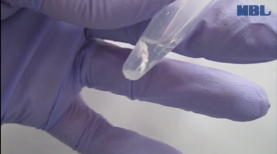
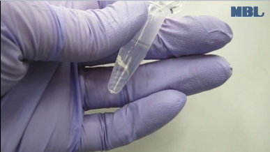
Incubation with a primary antibody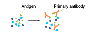 Add 500 µL of protein extract and 2-10 µg of the primary antibody to a 1.5-mL tube. |
 |
| Incubate at 4°C for 1 hour-overnight with shaking on a rotator. | 
|
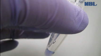
Incubation with a secondary antibody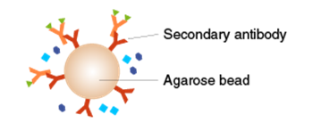 Add secondary antibody-coated (or protein A/G-coated) agarose beads. |
 |
| Incubate at 4°C for 1 hour-overnight on a rotating shaker. | |
Washing Pellet the agarose beads by centrifugation, and remove the supernatant by aspiration without disturbing the beads. |
|
| Add 1 mL of ice-cold lysis buffer or washing buffer, mix, and centrifuge. Remove the supernatant by aspiration without disturbing the beads. Repeat 3-4 times. |
|
| <When performing SDS-PAGE or Western blotting> Add 50 µL of 2x SDS sample buffer containing 2-mercaptoethanol (2-ME) and heat for 5 minutes to elute the target protein from the beads. Centrifuge, and use the supernatant for SDS-PAGE.  ※The antibodies are co-eluted, which should be taken into account when analyzing the data. |
|
| <When extracting the target protein while preserving its activity and native conformation (pull-down assay)> Elute the target protein in neutral pH using an elution peptide and avoid harsh conditions, such as acidic and alkaline solutions. [Related topics] Pull-down assay experiments using Tagged protein purification kits |
|
>>The principle and method of Western blotting (WB)
>>The principle and method of polyacrylamide gel electrophoresis (SDS-PAGE)