What are Organoids?
What are Organoids?
Organoids are miniaturized organs that are derived from stem cells in vitro. They are formed as three-dimensional tissue-like structures by utilizing the self-renewal and multi-differentiation capacities of stem cells.

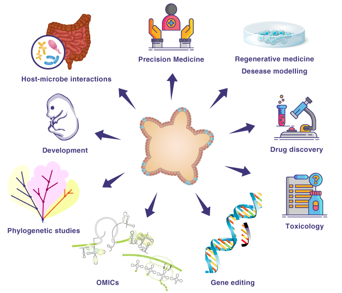
Compared to conventional cultured cells and spheroids, organoids are anatomically and functionally close to the organs in the living body. Organoids can be constructed from both normal and diseased tissues, allowing for utilization in various research applications. These tissue structures are used for basic research such as embryology, physiology, and evolution as well as more involved research including characterizing pathological disease conditions and drug discovery research. In addition, there are interesting studies using applied research for regenerative medicine using organoids for transplantation.
Types and Applications of Organoids
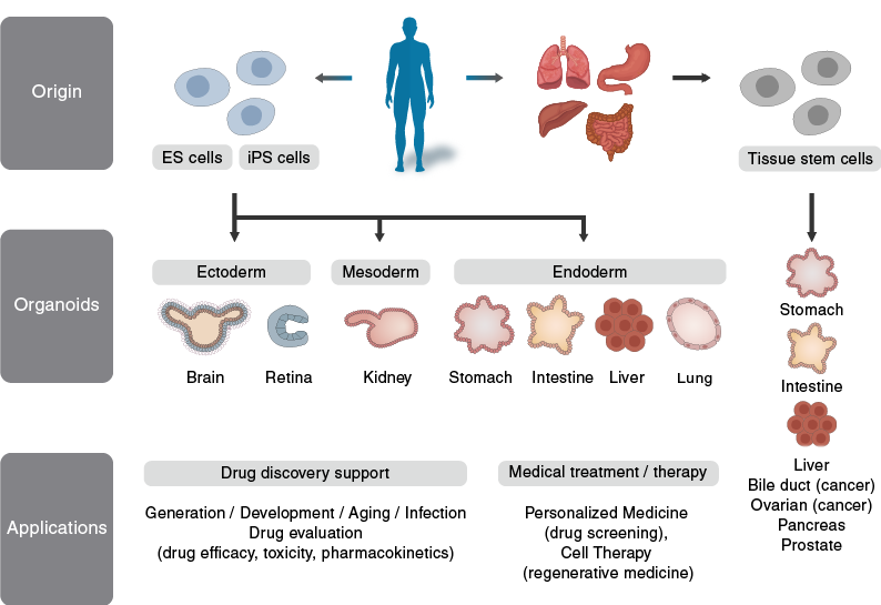
Organoids are mainly derived from pluripotent stem cells (iPS cells, ES cells) and organoids derived from tissue stem cells.

Utilization of organoid technology
Organoid technology is utilized in research areas such as embryology and the investigation of disease mechanisms. Organoids also used in drug discovery research for applications such as drug screening and regenerative medicine. The use of organoids has allowed for important downstream discoveries for better physiological understanding in order to improve human health and body.
Schematic diagram of organoid technology

Applications of pluripotent stem cell organoids
Research on organoids derived from pluripotent stem cells has elucidated the differentiation mechanism of each organ, leading to the investigation of disease mechanisms, drug screening, and application to regenerative medicine.
Table 4
human iPSC-derived organoids for modeling development and disease
| Starting cells | Organoids | Applications | References |
|---|---|---|---|
| Human ESCs and iPSCs | Cerebral organoids | Modeling human cortical development and microcephaly | Lancaster et al. 2013117 |
| Human ESCs and iPSCs | Cerebral organoids | Modeling human fetal neocortex development | Camp et al. 2015107 |
| Human iPSCs | Brain organoids | Modeling autism spectrum disorders | Mariani et al. 2015108 |
| Human ESCs and iPSCs | Brain organoids | Modeling Zika virus infection | Cugola et al. 2016109 |
| Human iPSCs | Brain-region-specific organoids | Modeling Zika virus infection. Future applications include modeling human brain development and diseases and drug testing. | Qian et al. 2016110 |
| Human iPSCs | Brain organoids | Modeling Zika virus infection | Garcez et al. 2016111 |
| Human iPSCs | Brain organoids | Modeling Seckel syndrome | Gabriel et al. 2016112 |
| Human ESCs and iPSCs | Cortical organoids | Modeling species difference | Otani et al. 2016113 |
| Human iPSCs | Cortical spheroids | Modeling cortical development and diseases | Pasca et al. 2015114 |
| Human iPSCs | Retinal organoids | Modeling glaucoma | Tucker et al. 2014106 |
| Human ESCs and iPSCs | Intestinal organoids | Studying intestinal development. Future applications include intestinal disease modeling. | Spence et al. 2011103 |
| Human ESCs and iPSCs | Small intestinal organoids | Transplantation to demonstrate responsiveness to systemic signals. Future applications include studying intestinal development and diseases. | Watson et al. 2014105 |
| Human iPSC | Liver bud | Organ-bud transplantation for regenerative medicine | Takebet et al. 201399 |
| Human iPSCs | Cystic organoids | Modeling Alagille syndrome, polycystic liver disease and cystic fibrosis-associated cholangiopathy and drug validation | Sampaziotis et al. 2015100 |
| Human iPSCs | Cystic organoids | Studying biliary development and cystic fibrosis | Ogawa et al. 2015101 |
| Human iPSC | Lung organoids | Studying lung development. Future applications include lung disease modeling. | Dye et al. 2015102 |
| Human iPSCs | Kidney organoids | Future applications include kidney disease modeling, nephrotoxicity screening and cell therapy. | Takasato et al. 2015104 |
| Human ESCs and iPSCs | Gastric organoids | Modeling gastric development and H. pylori infection | McCracken et al. 201698 |
Adapted from Table 4 of Yanhong Shi et al. “Induced pluripotent stem cell technology: a decade of progress”
Nat Rev Drug Discov. Vol. 16 no.2 (2017) : 115–130.


