Antibody detailed product information
| Code No. | M148-3M | |
| Anti-monomeric Kusabira-Green N-terminal fragment mAb | ||
| Size | 100 µL (1 mg/mL) | |
| Availability (in Japan) | 1-9
(In Japan at 17:33, Apr 26, 2024 in JST) |
|
| Clonality | Monoclonal | |
| Clone | 1E6 | |
| Isotype (Immunized Animal) |
Mouse IgG2b | |
| Applications | WB 1 µg/mL IC* (PMID: 22911572) |
|
| Immunogen (Antigen) |
mKG_N (1-168 a.a.) | |
| Storage buffer | 1 mg/mL in PBS/50% glycerol, pH 7.2 | |
| Storage temp. | -20°C | |
| Conjugate | Unlabeled | |
| Manufacturer | MBL | |
| Alternative names | mKG_N | |
| Background | CoralHue® Fluo-chase Kit can detect protein-protein interactions as fluorescent signals using the protein fragment complementation method. The gene of CoralHue® monomeric Kusabira-Green (mKG), a reporter protein, is divided into two fragments (CoralHue® mKG_N fragment and CoralHue® mKG_C fragment) which are respectively fused to the target protein genes to investigate the interactions. When the expressed target proteins don’t interact, CoralHue® mKG_N fragment and CoralHue® mKG_C fragment cannot approach each other and can not emit fluorescence. However, when target proteins interact, divided CoralHue® mKG fragments spatially approach each other and the local effective concentration increases. As a result, mKG fragments form a steric structure before dividing and the chromophore emits fluorescence. The fluorescent signals can be detected depending on the fused target protein-protein interactions. | |
| Related products | M149-3M Anti-monomeric Kusabira-Green C-terminal fragment mAb M168-3M Anti-monomeric Kusabira-Orange 2 mAb |
|
| Notes | This antibody (clone 1E6) has the epitope on CoralHue® mKG_N fragment and can detect the fusion protein with CoralHue® mKG_N specifically. | |
| Product category | Tools:Fluorescent proteins | |
| Data | 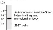 |
|
| Citations | Immunocytochemistry
|
|
- The availability is based on the information in Japan at 17:33, Apr 26, 2024 in JST.
- The special price is shown in red color.
- Please note that products cannot be ordered from this website. To purchase the items listed in this website, please contact us or local distributers.
- Abbreviations for applications:
WB: Western Blotting, IH: Immunohistochemistry, IC: Immunocytochemistry, IP: Immunoprecipitation
FCM: Flow Cytometry, NT: Neutralization, IF: Immunofluorescence, RIP: RNP Immunoprecipitation
ChIP: Chromatin Immunoprecipitation, CoIP: Co-Immunoprecipitation
DB: Dot Blotting, NB: Northern Blotting, RNA FISH: RNA Fluorescence in situ hybridization - For applications and reactivity:
*: The use is reported in a research article (Not tested by MBL). Please check the data sheet for detailed information.
**: The use is reported from the licenser (Under evaluation or not tested by MBL).
- For storage temparature: RT: room temparature
- Please note that products in this website might be changed or discontinued without notification in advance for quality improvement.